
T. Niki, S. Saito, and D. K. Gladish
 https://doi.org/10.1080/10520295.2019.1601769
ABSTRACT
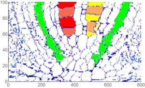
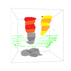
We developed a novel sectioning and staining method to make high contrast, high resolution sections of plant tissue for light microscopy. Specimens of teosinte (Zea mays L., ssp. mexicana) root tips were fixed and embedded in Technovit 7100™ plastic resin. Thin sections, 1−2.5 μm, were cut and mounted on glass slides. The sections were either treated with RNase or not, then stained with 0.1% toluidine blue O and observed through ∞/0 objective lenses. For light microscopy, the enzyme staining procedure increased resolution and contrast. High magnification ∞/0 objective lenses produced high quality images for digital photography without using a coverslip or immersion oil. Our slide preparation and microscopic analysis were less labor intensive and more rapid than previous methods and enabled rapid and precise alignment of serial transverse sections for both tracking cell lineages and tissue measurements.