This website is being redirected to the following URL.
https://www.mikion.org


ミキ音響
This website is being redirected to the following URL.
https://www.mikion.org
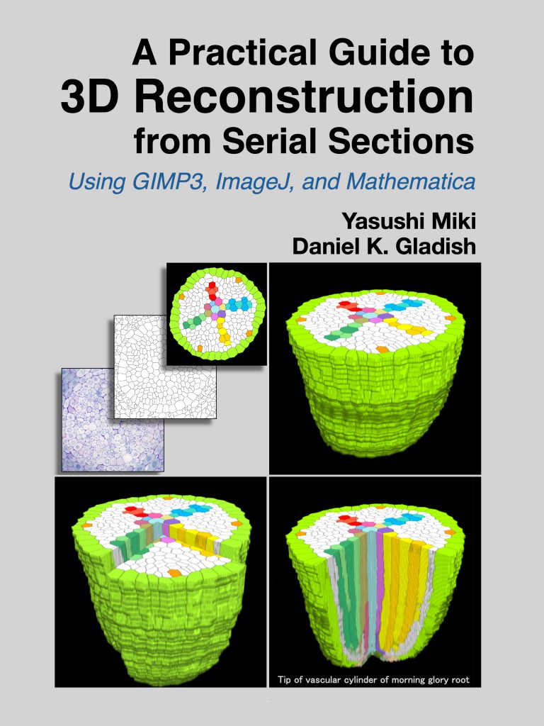
A Practical Guide to 3D Reconstruction from Serial Sections: Using GIMP3, ImageJ, and Mathematica
by Yasushi Miki (Author), Daniel K. Gladish (Author)
ISBN-13: 979-8268447194
Publication date: October 25, 2025
Kindle, Paperback
https://www.mikion.org
に移動処理進行中です.
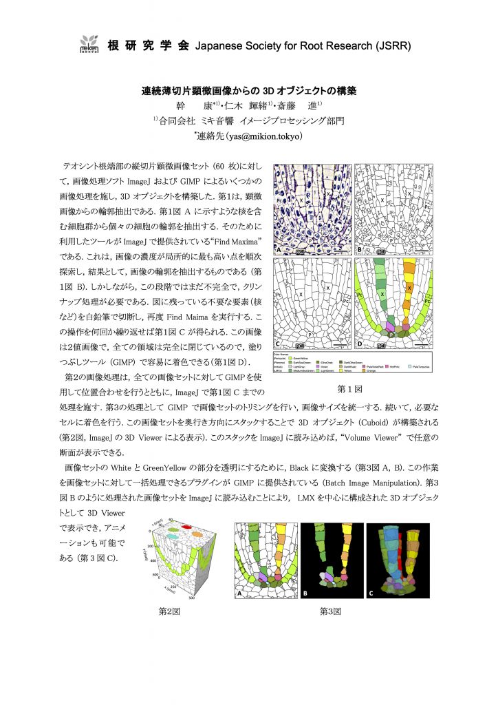
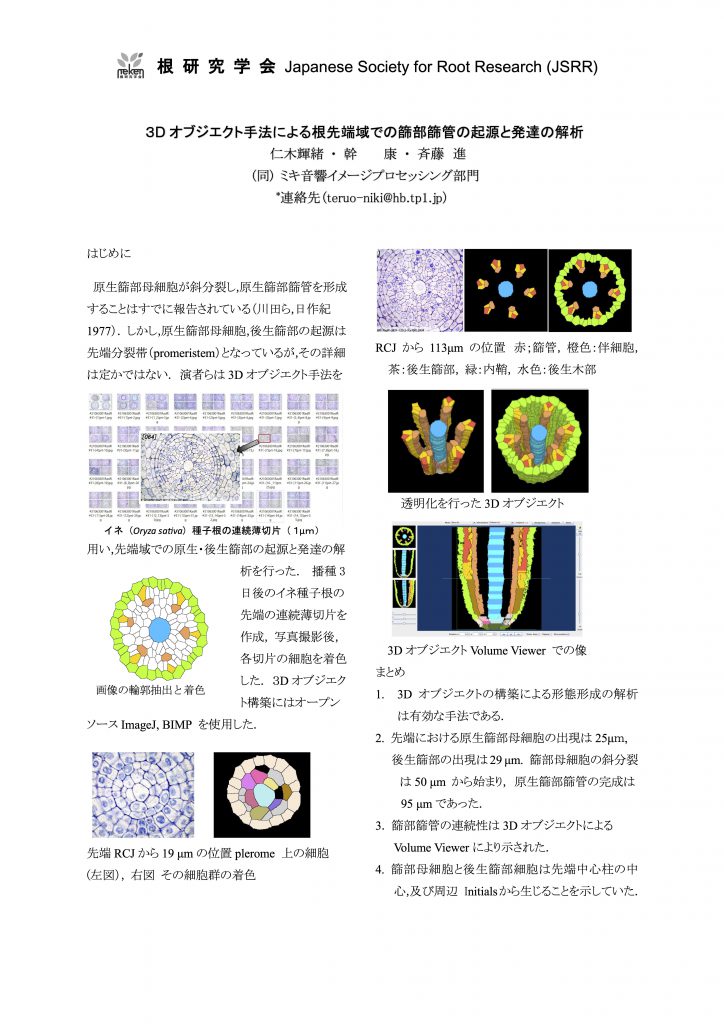
Yasushi Miki, Susumu Saito, Teruo Niki and Daniel K. Gladish
Plants 2024, 13(6), 910; https://doi.org/10.3390/plants13060910
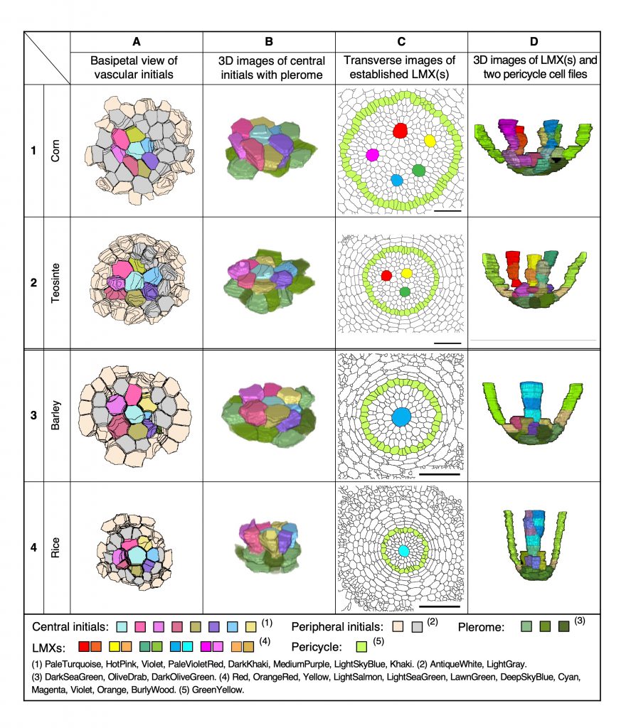
Figure 9. Characteristics of the primary root tip procambia of four taxa (1–4) in the Poaceae as seen in enhanced section images and 3D constructions. (A) Views of the basipetal face of the vascular initials layers, (B) oblique perspective of 3D constructions of the central initials and plerome cells (shades of green), (C) transverse images of the VC at 100 µm from the RCJ, and (D) 3D images from the primary root tips of the LMX(s) and two pericycle cell files associated with their plerome and vascular initials layer. Scale bar = 50 µm.
仁木輝緒 ・ 幹 康 ・ 斉藤 進
(同) ミキ音響イメージプロセッシング部門
第58回根研究集会(2023年11月3–5日,姫路)

Amazon公式サイトで「連続切片」で検索してください. オンデマンド(ペーパーバック)2023年7月25日発売 Kindle版2023年7月19日発売
Yasushi Miki, Susumu Saito, Teruo Niki and Daniel K. Gladish
Applications in Plant Sciences, vol. 11, July 2023
https://doi.org/10.1002/aps3.11531
幹 康・仁木輝緒・斉藤 進
ミキ音響イメージプロセッシング部門
第57回根研究集会(2023年5月20–21日,川崎)